
 说明书
说明书 MSDS
MSDS产品描述:Rabbit polyclonal antibody to HSPH1免疫原:KLH-conjugated synthetic peptide encompassing a sequence within the C-term region of human HSPH1. The exact sequence is proprietary.纯化方式:The antibody was purified by immunogen affinity chromatography.克隆类型:Polyclonal产品形式:Liquid in 0.42% Potassium phosphate, 0.87% Sodium chloride, pH 7.3, 30% glycerol, and 0.01% sodium azide.稀释比:WB (1/500 - 1/1000), IH (1/100 - 1/200), IF/IC (1/100 - 1/500)基因名称:HSPH1相关名称:HSP105; HSP110; KIAA0201; Heat shock protein 105 kDa; Antigen NY-CO-25; Heat shock 110 kDa protein
基因编号(人):
10808;
基因编号(小鼠):
15505;
基因编号(大鼠):
288444;
蛋白编号(人):
Q92598;
蛋白编号(小鼠):
Q61699;
蛋白编号(大鼠):
Q66HA8;
储存效期:Shipped at 4°C. Upon delivery aliquot and store at -20°C for one year. Avoid freeze/thaw cycles.
-
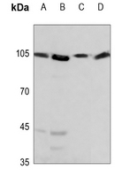 Western blot analysis of HSPH1 expression in A375 (A), DLD (B), mouse testis (C), rat testis (D) whole cell lysates. (Predicted band size: 96 kD; Observed band size: 105 kD)
 -
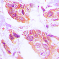 Immunohistochemical analysis of HSPH1 staining in human lung cancer formalin fixed paraffin embedded tissue section. The section was pre-treated using heat mediated antigen retrieval with sodium citrate buffer (pH 6.0). The section was then incubated with the antibody at room temperature and detected using an HRP conjugated compact polymer system. DAB was used as the chromogen. The section was then counterstained with haematoxylin and mounted with DPX.
 -
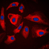 Immunofluorescent analysis of HSPH1 staining in HeLa cells. Formalin-fixed cells were permeabilized with 0.1% Triton X-100 in TBS for 5-10 minutes and blocked with 3% BSA-PBS for 30 minutes at room temperature. Cells were probed with the primary antibody in 3% BSA-PBS and incubated overnight at 4 °C in a humidified chamber. Cells were washed with PBST and incubated with a DyLight 594-conjugated secondary antibody (red) in PBS at room temperature in the dark. DAPI was used to stain the cell nuclei (blue).
 -
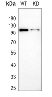 Western blot analysis of HSPH1 expression in wild type (WT) and knockdown (KD) HeLa cell lysates.