
 说明书
说明书 MSDS
MSDS产品描述:Rabbit polyclonal antibody to DOK4免疫原:Recombinant full length protein of human DOK4纯化方式:The antibody was purified by immunogen affinity chromatography.克隆类型:Polyclonal产品形式:Liquid in 0.42% Potassium phosphate, 0.87% Sodium chloride, pH 7.3, 30% glycerol, and 0.01% sodium azide.稀释比:WB (1/500 - 1/2000), IF/IC (1/50 - 1/200)基因名称:DOK4相关名称:Docking protein 4; Downstream of tyrosine kinase 4; Insulin receptor substrate 5; IRS-5; IRS5
基因编号(人):
55715;
基因编号(小鼠):
114255;
蛋白编号(人):
Q8TEW6;
蛋白编号(小鼠):
Q99KE3;
储存效期:Shipped at 4°C. Upon delivery aliquot and store at -20°C for one year. Avoid freeze/thaw cycles.
-
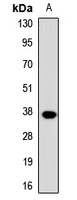 Western blot analysis of DOK4 expression in MCF7 (A) whole cell lysates. (Predicted band size: 37 kD; Observed band size: 37 kD)
 -
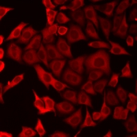 Immunofluorescent analysis of DOK4 staining in HeLa cells. Formalin-fixed cells were permeabilized with 0.1% Triton X-100 in TBS for 5-10 minutes and blocked with 3% BSA-PBS for 30 minutes at room temperature. Cells were probed with the primary antibody in 3% BSA-PBS and incubated overnight at 4 °C in a humidified chamber. Cells were washed with PBST and incubated with a DyLight 594-conjugated secondary antibody (red) in PBS at room temperature in the dark.