
 说明书
说明书 MSDS
MSDS产品描述:Rabbit polyclonal antibody to LC3A/B免疫原:Recombinant full length protein of human LC3A/B纯化方式:The antibody was purified by immunogen affinity chromatography.克隆类型:Polyclonal产品形式:Liquid in 0.42% Potassium phosphate, 0.87% Sodium chloride, pH 7.3, 30% glycerol, and 0.01% sodium azide.稀释比:WB (1/500 - 1/2000), IH (1/50 - 1/200), IF/IC (1/50 - 1/200)基因名称:MAP1LC3A; MAP1LC3B相关名称:Microtubule-associated proteins 1A/1B light chain 3A; Autophagy-related protein LC3 A; Autophagy-related ubiquitin-like modifier LC3 A; MAP1 light chain 3-like protein 1; MAP1A/MAP1B light chain 3 A; MAP1A/MAP1B LC3 A; Microtubule-associated protein 1 light chain 3 alpha; MAP1ALC3; Microtubule-associated proteins 1A/1B light chain 3B; Autophagy-related protein LC3 B; Autophagy-related ubiquitin-like modifier LC3 B; MAP1 light chain 3-like protein 2; MAP1A/MAP1B light chain 3 B; MAP1A/MAP1B LC3 B; Microtubule-associated protein 1 light chain 3 beta
基因编号(人):
84557;
81631;
基因编号(小鼠):
66734;
67443;
基因编号(大鼠):
362245;
64862;
蛋白编号(人):
Q9H492;
Q9GZQ8;
蛋白编号(小鼠):
Q91VR7;
Q9CQV6;
蛋白编号(大鼠):
Q6XVN8;
Q62625;
储存效期:Shipped at 4°C. Upon delivery aliquot and store at -20°C for one year. Avoid freeze/thaw cycles.
-
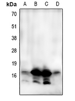 Western blot analysis of LC3A/B expression in SHSY5Y (A), HepG2 (B), BT474 (C), mouse brain (D) whole cell lysates. (Predicted band size: 14 kD; Observed band size: 14; 17 kD)
 -
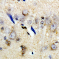 Immunohistochemical analysis of LC3A/B staining in human brain formalin fixed paraffin embedded tissue section. The section was pre-treated using heat mediated antigen retrieval with sodium citrate buffer (pH 6.0). The section was then incubated with the antibody at room temperature and detected using an HRP conjugated compact polymer system. DAB was used as the chromogen. The section was then counterstained with haematoxylin and mounted with DPX.
 -
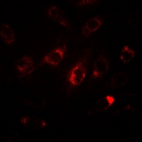 Immunofluorescent analysis of LC3A/B staining in HepG2 cells. Formalin-fixed cells were permeabilized with 0.1% Triton X-100 in TBS for 5-10 minutes and blocked with 3% BSA-PBS for 30 minutes at room temperature. Cells were probed with the primary antibody in 3% BSA-PBS and incubated overnight at 4 °C in a humidified chamber. Cells were washed with PBST and incubated with a DyLight 594-conjugated secondary antibody (red) in PBS at room temperature in the dark.