
 说明书
说明书 MSDS
MSDS产品描述:Mouse monoclonal antibody to Cytokeratin 18免疫原:Recombinant protein corresponding to human Cytokeratin 18.纯化方式:Affinity chromatography克隆类型:Monoclonal产品形式:Liquid in 0.42% Potassium phosphate, 0.87% Sodium chloride, pH 7.3, 30% glycerol, and 0.01% sodium azide.稀释比:WB (1/1000 - 1/3000), IF/IC (1/100 - 1/200)基因名称:KRT18相关名称:CYK18; Keratin type I cytoskeletal 18; Cell proliferation-inducing gene 46 protein; Cytokeratin-18; CK-18; Keratin-18; K18
基因编号(人):
3875;
基因编号(小鼠):
16668;
基因编号(大鼠):
294853;
蛋白编号(人):
P05783;
蛋白编号(小鼠):
P05784;
蛋白编号(大鼠):
Q5BJY9;
储存效期:Shipped at 4°C. Upon delivery aliquot and store at -20°C for one year. Avoid freeze/thaw cycles.
-
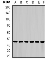 Western blot analysis of Cytokeratin 18 expression in HepG2 (A), Hela (B), mouse liver (C), mouse skeletal muscle (D), C2C12 (E), rat heart (F) whole cell lysates. (Predicted band size: 48 kD; Observed band size: 46 kD)
 -
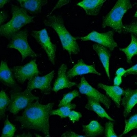 Immunofluorescent analysis of Cytokeratin 18 staining in Hela cells. Formalin-fixed cells were permeabilized with 0.1% Triton X-100 in TBS for 5-10 minutes and blocked with 3% BSA-PBS for 30 minutes at room temperature. Cells were probed with the primary antibody in 3% BSA-PBS and incubated overnight at 4 °C in a hidified chamber. Cells were washed with PBST and incubated with a FITC-conjugated secondary antibody (green) in PBS at room temperature in the dark. DAPI was used to stain the cell nuclei (blue).