
 说明书
说明书 MSDS
MSDS产品描述:Rabbit polyclonal antibody to Histone H3 (TriMethyl-K27)免疫原:KLH-conjugated synthetic Tri-methylated peptide corresponding to residues surrounding K27 of human Histone H3 protein. The exact sequence is proprietary.纯化方式:The antibody was purified by immunogen affinity chromatography.克隆类型:Polyclonal产品形式:Liquid in 0.42% Potassium phosphate, 0.87% Sodium chloride, pH 7.3, 30% glycerol, and 0.01% sodium azide.稀释比:WB (1/500 - 1/1000), IH (1/50 - 1/200), IF/IC (1/50 - 1/200), ChIP (1/10 - 1/50)基因名称:HIST1H3A; HIST1H3; HIST1H3C; HIST1H3D; HIST1H3E; HIST1H3F; HIST1H3G; HIST1H3H; HIST1H3I; HIST1H3J; HIST2H3A; HIST2H3C; HIST2H3D; H3F3A; H3F3B; H3F3C相关名称:HIST1H3A; H3FA; HIST1H3B; H3FL; HIST1H3C; H3FC; HIST1H3D; H3FB; HIST1H3E; H3FD; HIST1H3F; H3FI; HIST1H3G; H3FH; HIST1H3H; H3FK; HIST1H3I; H3FF; HIST1H3J; H3FJ; Histone H3.1; Histone H3/a; Histone H3/b; Histone H3/c; Histone H3/d; Histone H3/f; Histone H3/h; Histone H3/i; Histone H3/j; Histone H3/k; Histone H3/l; HIST2H3A; HIST2H3C; H3F2; H3FM; HIST2H3D; Histone H3.2; Histone H3/m; Histone H3/o; H3F3A; H3.3A; H3F3; PP781; H3F3B; H3.3B; Histone H3.3; H3F3C; Histone H3.3C; Histone H3.5
基因编号(人):
8350;
8351;
8352;
8353;
8354;
8355;
8356;
8357;
8358;
8968;
126961;
333932;
653604;
3020;
3021;
440093;
蛋白编号(人):
P68431;
Q71DI3;
P84243;
Q6NXT2;
储存效期:Shipped at 4°C. Upon delivery aliquot and store at -20°C for one year. Avoid freeze/thaw cycles.
-
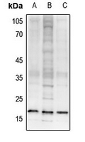 Western blot analysis of Histone H3 (TriMethyl-K27) expression in SHSY5Y (A), C6 (B), 3T3L1 (C) whole cell lysates. (Predicted band size: 15 kD; Observed band size: 17 kD)
 -
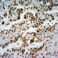 Immunohistochemical analysis of Histone H3 (TriMethyl-K27) staining in human lung cancer formalin fixed paraffin embedded tissue section. The section was pre-treated using heat mediated antigen retrieval with sodium citrate buffer (pH 6.0). The section was then incubated with the antibody at room temperature and detected using an HRP conjugated compact polymer system. DAB was used as the chromogen. The section was then counterstained with haematoxylin and mounted with DPX.
 -
 Immunofluorescent analysis of Histone H3 (TriMethyl-K27) staining in C6 cells. Formalin-fixed cells were permeabilized with 0.1% Triton X-100 in TBS for 5-10 minutes and blocked with 3% BSA-PBS for 30 minutes at room temperature. Cells were probed with the primary antibody in 3% BSA-PBS and incubated overnight at 4 °C in a hidified chamber. Cells were washed with PBST and incubated with an AF488-conjugated secondary antibody (green) in PBS at room temperature in the dark. Phalloidin - AF594 was used to stain Actin filaments (red). DAPI was used to stain the cell nuclei (blue).
 -
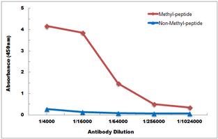 Direct ELISA antibody dose-response curve using Anti-Histone H3 (TriMethyl-K27) Antibody. Antigen (methyl-peptide and non-methyl-peptide) concentration is 5 ug/ml. Goat Anti-Rabbit IgG (H&L) - HRP was used as the secondary antibody, and signal was developed by TMB substrate.